1 Introduction
This document will describe the chemistry of life, from how the atom works, right up to cells,
then viruses and finally vaccines.
Lastly there is a chapter on how we know all this stuff, how scientists work it out.
I will use the least amount of technical terms, and when I do use one, I will describe
it first.
I will also use some terms of my own. The first I will tell here, and that is
“soup”.
I use the word soup to describe a place where there is an abundance of everything.
By everything I mean atoms, molecules and stuff. Examples of soup are, the ocean,
the atmosphere, the blood system and most importantly the inside of human cells.
When describing a chemical reaction we often say “and a water molecule does…”,
well where did that water molecule come from? Well it was just in the soup,
floating around at the time.
This text attempts to make everything simple, and so sometimes I do not use a complete
technical description. You should be able to read the entire thing in about an hour, and
hopefully come out at the end with a better understanding of what viruses are.
Because of the COVID-19 pandemic, I shall use the coronavirus a lot, but everything
described applies to all viruses.
If you know what each chapter is about, just click on the link in the contents to
jump forward.
I describe everything from scratch for completeness, and so you can find everything
you need.
And, so first, to the chemistry of life…
2 Atoms
The classic description of the atom is like a solar system, with the planets whizzing
around the sun, here we shall say the electrons are whizzing around the nucleus.
Quantum theory describes the electrons as a probability field, but we shall ignore that here.
ASIDE: There are two definitions of nucleus in this text, one is the centre of the atom,
the other is the centre of the cell. They share the word, but are completely different things.
The main feature of ALL chemistry is the charge. Properly called the electrostatic charge,
it is a force of the universe that makes electrons be attracted to protons. Electrons have
minus (-) charge, and protons have plus (+) charge. And like bar magnets you played with at
school, when you pushed the two norths together and felt them repel, and then felt the
click of north and south snapping together by attraction, charge has a similar effect.
So, electrons repel each other, but are attracted to the proton.
The atom nucleus is made of protons and neutrons bound together. I will not describe the
forces (strong and weak nuclear) that do this, and I will ignore the neutral neutrons (hehe).
So protons are the nucleus. Now, imagine you have a nucleus in your hand the size of a pea,
and walk out to the middle of a football field, well the electrons would be whizzing around
on the boundary, and be the size of a pin prick.
Why do the electrons not rush in to the nucleus, well they are moving too fast. That is a
rule of the universe, that they never stop moving.
And the width of the atom is about 10-10 metre, so that means
there are 1,000,000 atoms in the
width of a human hair. Now as the atom is smaller than the wavelength of light, we can not
see it in an ordinary microscope no matter how much we magnify it.
There are just over 100 elements in the universe, and we list them in the Periodic Table
of Elements. Its sequence is by Atomic Number. This number is also the same as the number of
electrons, and as the number of protons. The first element is hydrogen,
with one proton and one electron.
It is amazing that life uses only three elements, Hydrogen, Oxygen and Carbon (H, O, C).
Well that is 99.8%, with 0.1% being Sulfur, Phosphorus and Nitrogen (S, P, N),
and then there about ten called the trace elements. All the rest of the elements are toxic to
life, like lead and mercury.
STRUCTURE
So, how does the chemistry work? Well it is all due to the outside shell (ring) of electrons.
Unlike the solar system analogy, electrons can group in shells, but only according to set
rules.
A shell can fill up with electrons, and then the next atomic number must start a new shell.
The maximum number of electrons that can exist in each shell are:
2 8 8 18 18 32
But since we are talking about the life elements, we will only look at the first two shells
(2 8). For life elements the electrons are Hydrogen=1, Carbon=6 and Oxygen=8.
ELEMENT
FIRST SHELL
SECOND SHELL
EMPTY HOLES
Hydrogen
1
0
1 (in the first shell)
Carbon
2
4
4 (in the second shell)
Oxygen
2
6
2 (in the second shell)
3 Molecules
Atoms that have holes in their outer shell like to bond with other atoms in the soup.
What happens is, that if an atom gets close enough to another atom in the soup they will bond.
They have to get really close, much less than 1/10th of the atom width.
So, an atom, like hydrogen that has a spare electron in the outer shell will share an electron
with an atom that has a free hole in its outer shell.
This is called a covalent bond, and is the strongest and most common bond.
There is also a hydrogen bond. This is where hydrogen bonds with Fluorine, Oxygen or
Nitrogen, only. We will deal with H to O bonds only. Hydrogen bonds are weaker than
covalent bonds.
So, we all know water is H2O, this looks like this in diagram form.
The straight lines are the bonds.
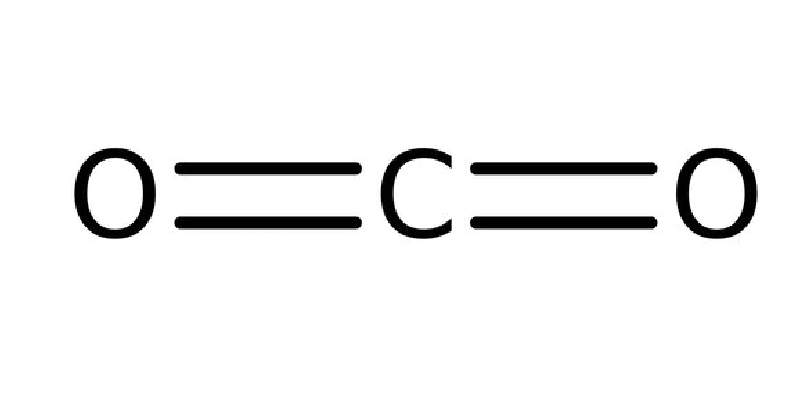
And carbon dioxide is CO2, which looks like this in diagram form.
Since Carbon has 4 holes, it has made a double bond
with each of the Oxygens.
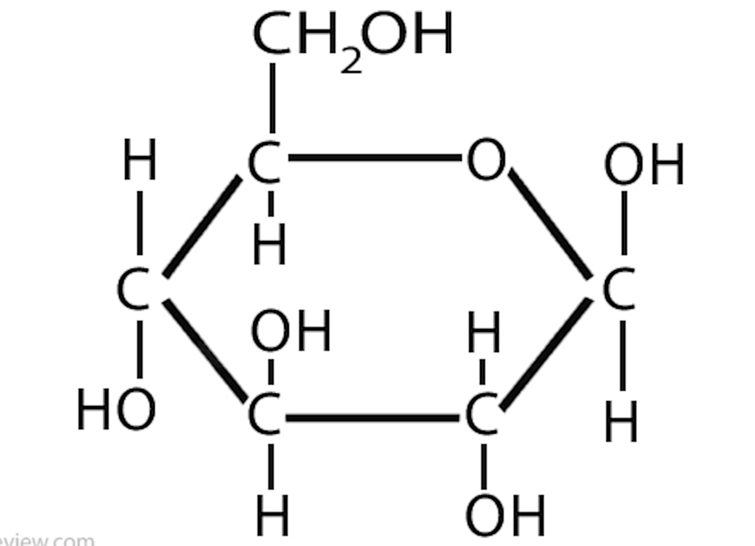
Glucose is C6H12O6 and below is the full diagram for glucose:
It gets a little complicated, so we drop the Carbon C letter, and all the corners are
now Carbon. We also know Carbon has 4 holes, so any bonds that are not shown will be hidden
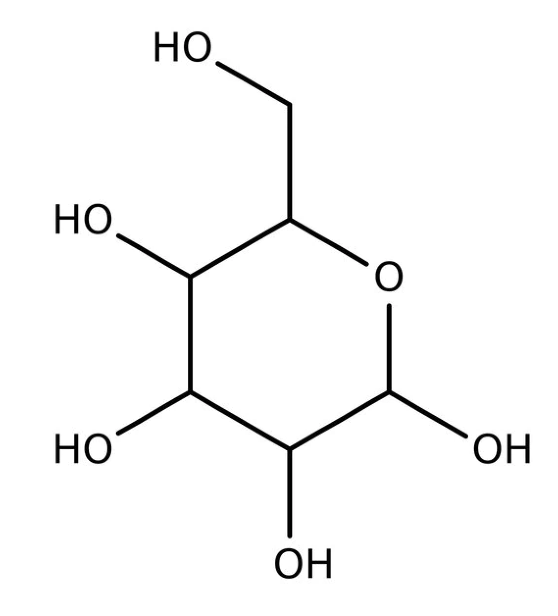
hydrogen. So a simplified diagram of glucose is:
There are 6 carbons, but no C letters, but count the corners.
And there are 6 Oxygens shown.
Also there are 7 hidden Hydrogens and 5 shown, making the 12 Hydrogens in Glucose.
We will see diagrams like these again later.
You do not need to know them all, I just show
them as a picture so you can see the molecules as they get more complex.
You see glucose is a basic hexagon shape. All life is based around these sugars, and we will
see the hexagon again.
4 Cells
I will go into a bit of detail about the cell, because it is the basis of life.
Cells are around 10-6 metre wide, and can be seen in a light microscope.
However some human cells like the sciatic nerve which goes from the spine all the way
down the leg, can be a meter long.
The largest cell in biology is the ostrich egg, which can be up to 1.5 kg.
The basis of the cell is the cell wall, also called the membrane.
The cell membrane is what keeps the contents away from the outside world.
The cell contents are where the chemistry of life mostly happens, and it needs protection.
CELL WALL
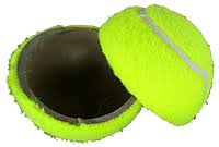
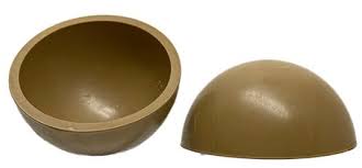
Think of a tennis ball, and cut it in half.
In simplified form it looks like a sphere cut in half.
So you can see the spherical wall, which kept the hollow centre separate from
the outside world. Now not all cells are spheres, but this idea will do for now.
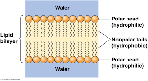
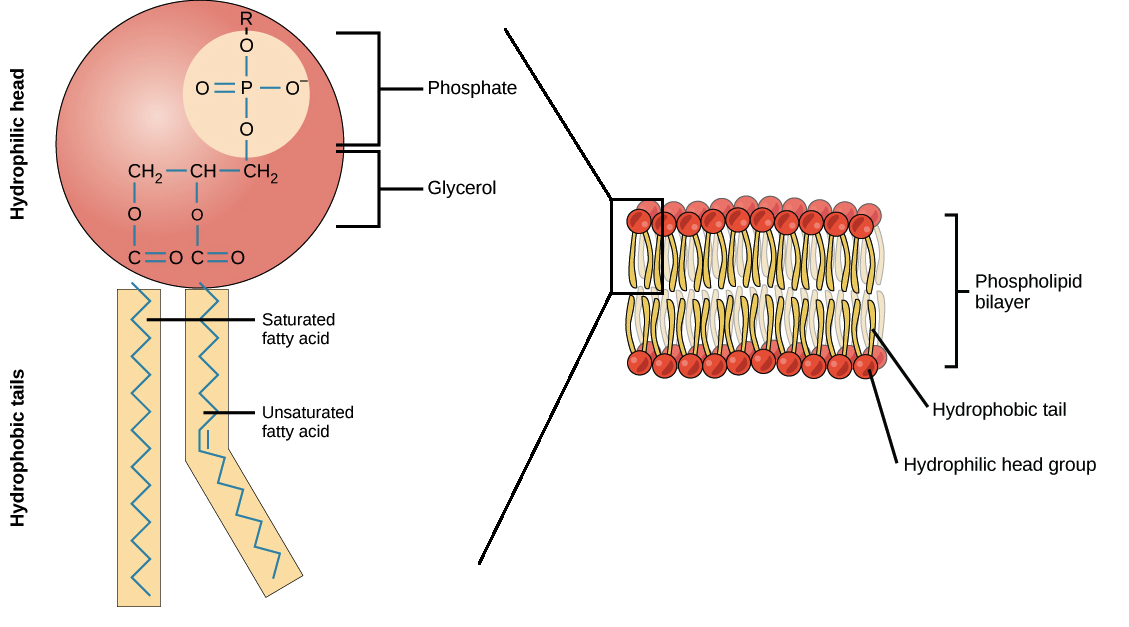
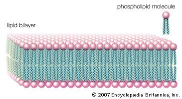
The cell wall is made of a lipid bi-layer…whew, what is that?
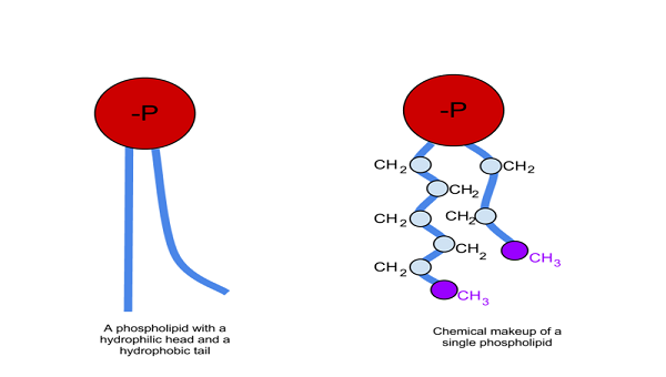
Lipids are fat molecules. No, not adipose cells like the fat on my belly.
They are a special molecule, and they look like this: There is a head, and two legs. The head contains the first life element which is
not H O C, and that is Phosphorus (P).
You can see the legs are a string of Carbons in the zig-zag Z shape.
The head likes water, and the legs do not like water, so they tend to clump together,
with the heads on the outside and the legs on the inside (hiding from the water).
And this layer forms into a sphere like the tennis ball. This is the cell membrane.
It is vital in our story, and we will revisit it many times.
The membrane is not impermeable, and many small molecules easily pass through, like water
and some gases.
Larger molecules however cannot pass through easily, and need to be invited in.
Also, the outside of the membrane is not smooth, and it interacts with the outside soup a
lot, with many molecules bonding to the outside. Many large molecules, such as enzymes and
proteins need to enter the cell, and they are invited in and gain entry by interacting with
molecules on the outside of the cell called receptors. We will meet receptors again later.
CELL CONTENTS
So we have our hollow sphere, and it is the inside where life happens. It is another soup,
and it is constantly active, and moving.
The cell shape is not really a sphere, and the actual cell shape is determined by millions
of tiny connectors called micro-tubules, which criss-cross the inside of the cell,
joining opposite sides of the membrane, giving the cell shape.
These microtubules are also the transport system of the cell, allowing the movement
of resources around the cell. We will meet microtubules again.
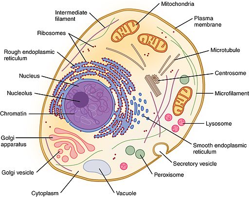
Also inside the cell are structures called organelles.
The most famous organelle is the nucleus.
There are also mitochondria, the endoplastic reticulum, and (my favourite) the
Golgi Apparatus, and many others. What lovely names!
We will only focus on the nucleus, as that is where DNA lives, and the story of DNA is
what comes next.
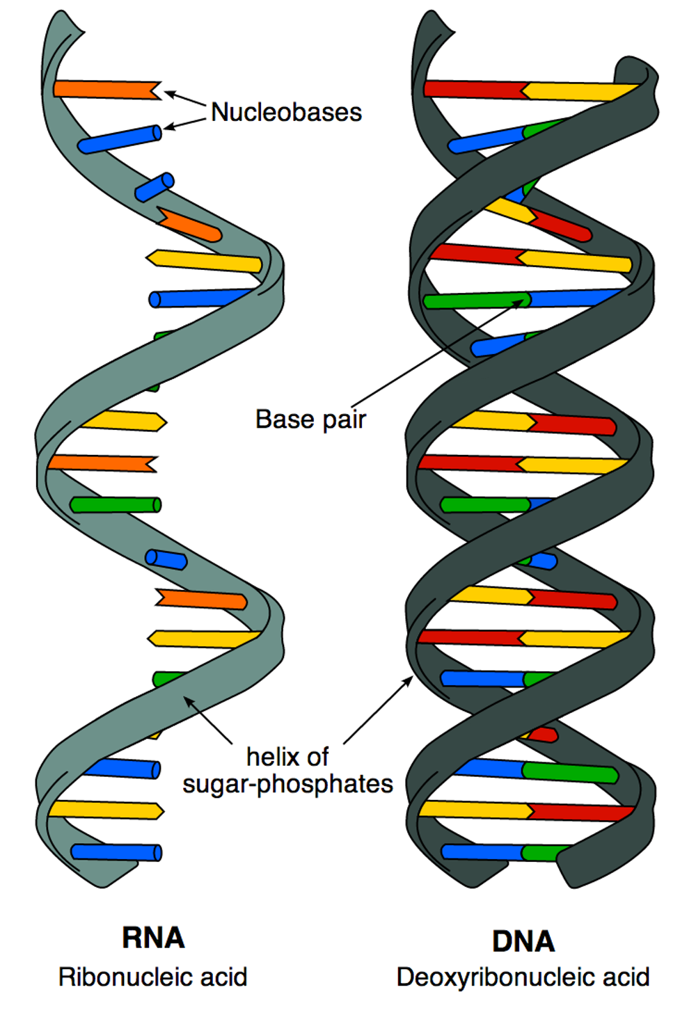
5 DNA
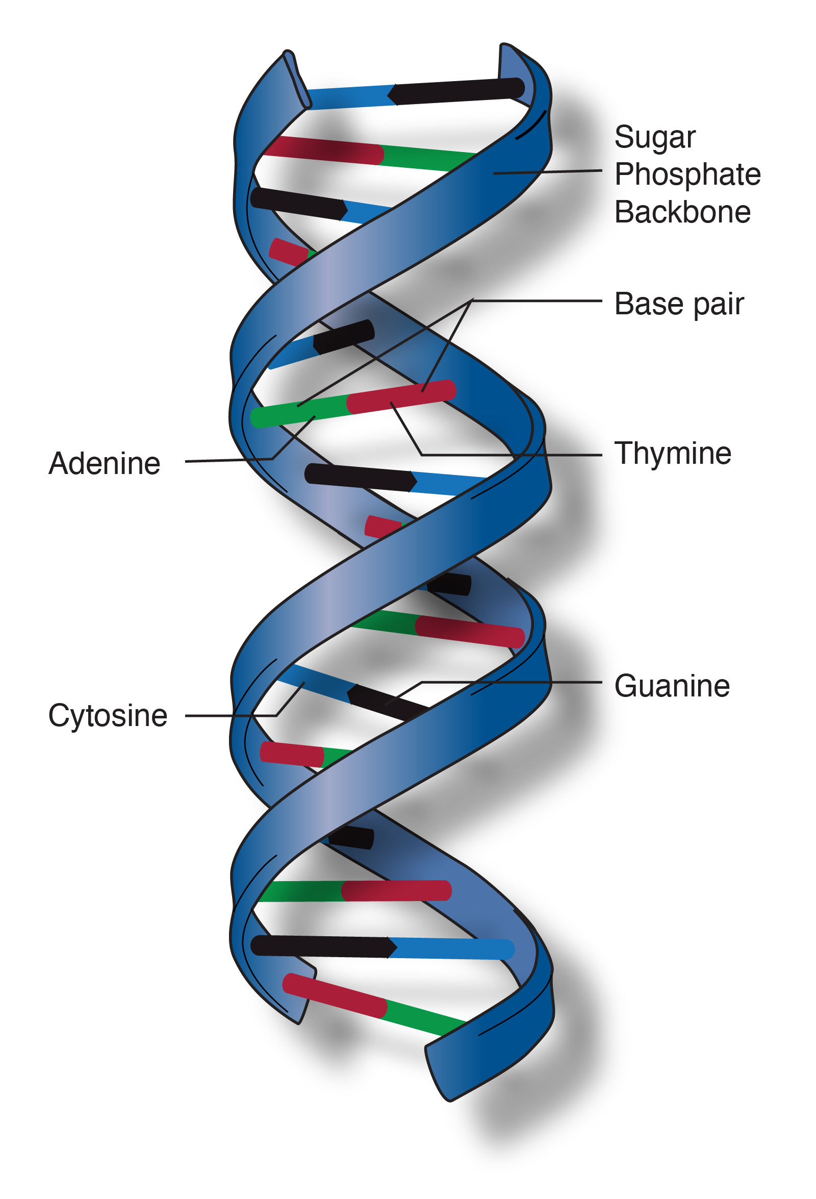
The correct term for DNA is DeoxyriboNucleic Acid, so let‘s just call it DNA.
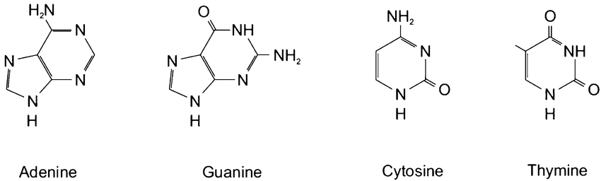
It is made up of a backbone (sugar phosphate) and four molecules C A T G, called nucleotides:
C - cytosine
A - adenine
T - thymine
G - guanine
OK so we have DNA and C A T G, we don‘t need to remember the long
technical terms. C A T G are also called bases.
Now take two combs and hold one in each hand. You are holding the backbone. Each prong on
the combs is a base. Hold the combs near each other with the prongs touching. They are
bonding, actually hydrogen bonds…remember those? Each prong is one of the C A T G, which
we call a base. Two prongs touching is a base-pair.
Now open the window, and extend the combs across the room, out the window, keep going
right over the horizon. The human DNA has 3,000,000,000 (3 billion base-pairs).
Now start to twist the joined combs, and do that a few million
times. You are making the classic DNA double helix (helix is just a spiral, like a spiral
staircase).
Now each time there is a C in one comb, there is a G in the other. This is a base pair.
And each time there is a T in one comb, there is an A in the other. So base pairs are
always A T, and C G. And also the reverse is true, T A, and G C. One comb is called the
strand, and the other comb is called the complimentary strand. So the sequence CATG has a
complimentary strand of GTAC.
So we have the backbone, the bases (hydrogen bonded), and it is twisted. This makes for a
strong molecule.
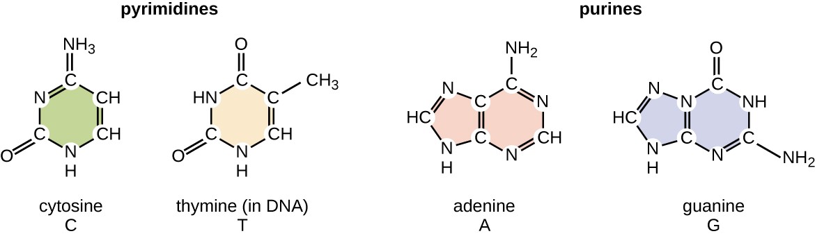
For completeness here are the diagrams for the C A T G. First the full diagram with the C
and all H showing:
And now simplified with the C missing, and the hidden H missing.
Also note the double bonds (like we had with carbon dioxide, earlier).
And here we revisit the hexagon sugar, and another pentagon (5-sided) structure.
We also introduce a new life element, Nitrogen (N).
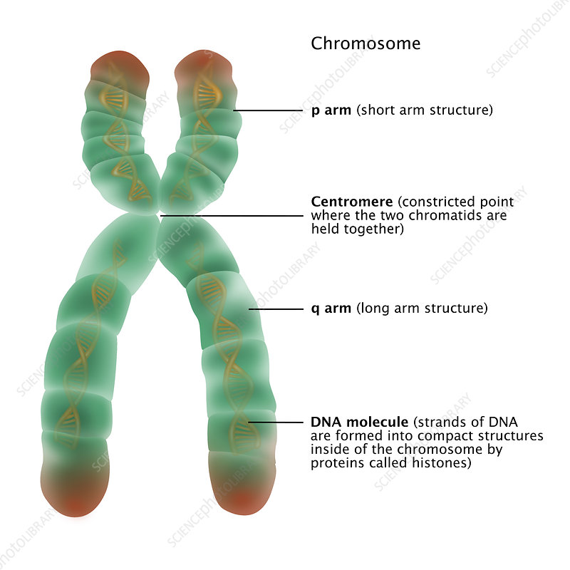
CHROMOSOMES
Now, like I said the DNA is a twisted helix. And anyone who ever played with a rope, or
cotton when sewing, will know that if you twist it, it will coil up around itself. And DNA
does exactly that. It coils up around itself, and then those coils coil further, and
eventually they end up in the shape of a chromosome, the classic shape that looks like a
stickman, with two arms and two legs.
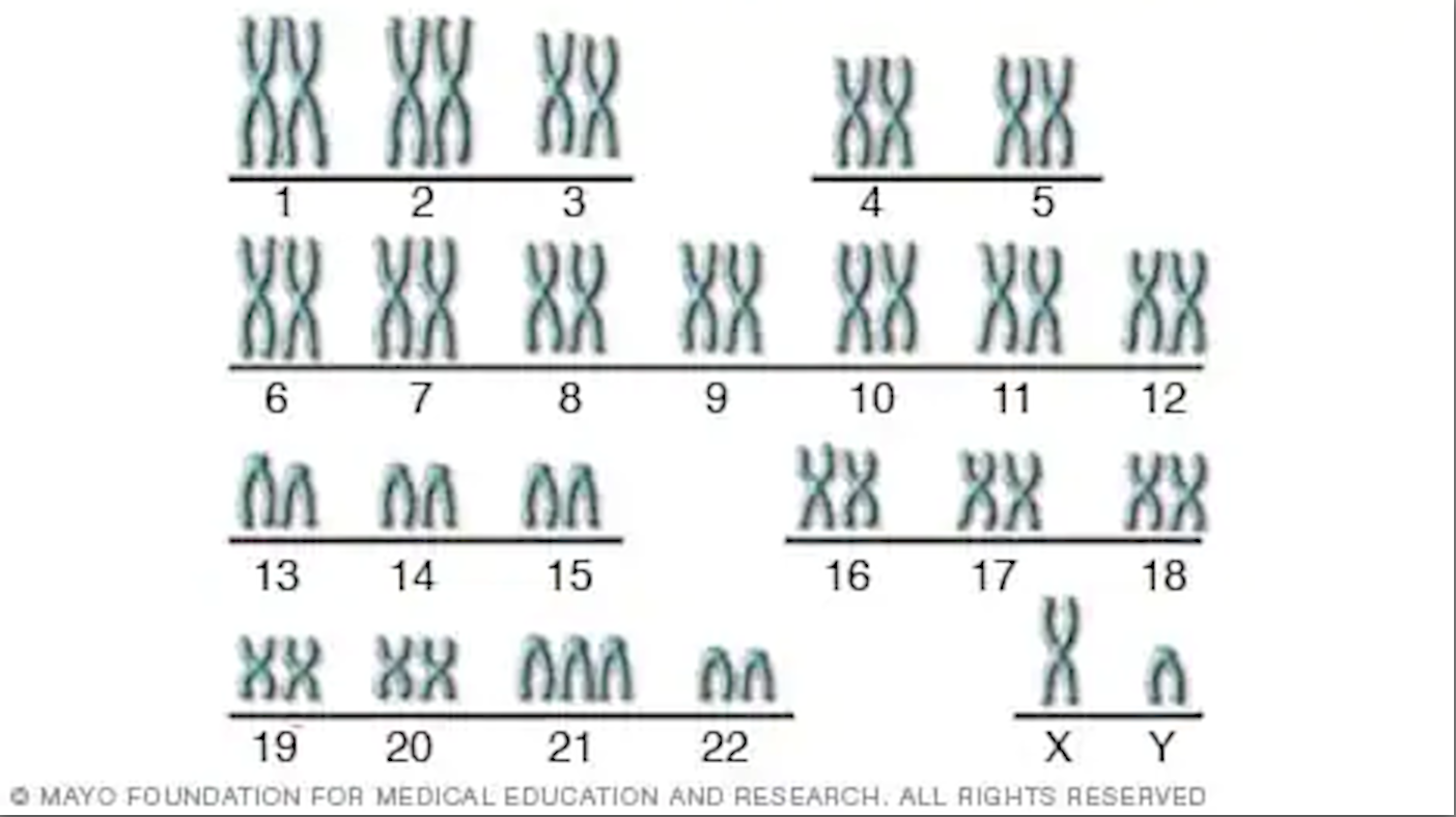
Every cell in your body has DNA in it. Well actually the red blood cells have no nucleus
so no DNA there. And the gametes, sperm and egg cells, only have half. The gametes have 22
chromosomes each, and one sex chromosome.
Chromosomes are categorised by their shape. Some have long arms, some long legs, and some
short versions. They are numbered by their shape. The 23rd chromosome is called the sex
chromosome, and comes in two shapes. One is a small X shape, and one is a small Y shape.
During conception, the sperm seeks out an egg, and when it finds it, it enters the egg.
The egg has a set of 23 chromosomes, and so does the sperm, and now there are 46 chromosomes
in the egg, and it is considered fertilised.
All egg cells only have an X shaped 23rd chromosome. Sperm cells can have either a small X,
or a small Y, 23rd chromosome. After fertilisation a cell with two X with become a girl,
and a cell with one X and one Y, will become a boy. So the sperm determines what gender
the baby will be.
So, girls have XX, and boys have XY sex chromosomes. Soon after fertilisation in the girl
egg cell, one of the X chromosomes will de-activate. How this is selected is currently
unknown.
CELL DIVISION
Soon after fertilisation the first cell division occurs. Technically this is called mitosis.
And mitosis continues during the whole pregnancy, and the baby will end up with thirty
trillion (30,000,000,000,000) cells, when she becomes an adult.
The first cells are stem cells, that is they are un-differentiated, but soon the DNA
stamps its authority on the foetus, and starts differentiating the cells into liver cells,
bone cells, all the different cells that make a body.
The DNA is the architect’s plan for the body. And since half the DNA comes from each parent,
the baby is the sum of the parents. Also since each egg and each sperm are slightly different,
you are slightly different to your brother or sister. Except for identical twins. At
conception it can happen that the first mitosis produces two babies, and so identical twins
have identical DNA. So, the DNA defines hair colour, eye colour and all the features of the
new human.
All through a persons life cells die. In fact apart from nerve cells, your entire body is
replaced every seven years. Some cells die from trauma, some from programmed cell death
(apoptosis). But for most of your adult life the cells that die are replaced by mitosis.
The process of mitosis is still being researched but the basic function is as follows.
The cell receives a signal to divide. The chromosomes line up down the centre of the cell,
and they all unzip. That is, the hydrogen bonds that held the base pairs together, separate.
So now there are the main strand and the complimentary strand separated, like two new combs
with their prongs sticking up. And for each base exposed a complimentary base in the soup
arrives and binds to it - A gets T, and C gets G. The backbone is also assembled, and when
completed (it takes about 20 minutes) there are now two sets of chromosomes where before
there was only one. They then move apart, a full set of 46 move to one side of the cell,
and the other 46 move to the opposite side of the cell. The cell membrane changes shape from
a basic “O” to an “8” isolating each set of chromosomes in a different lobe. And finally
the membrane separates into two, and mitosis is complete, and there are now two cells where
previously there was only one. All cells except blood cells, gametes and nerve cells do this
regularly.
During pregnancy DNA has the job of building a new body. After birth DNA has an ongoing
job of providing the body with the army of proteins it needs to keep the host alive.
6 Proteins
If you do an internet search on the word “protein” you will get millions of hits about how
much protein you need in your diet, which foods have the most protein and that they are
mostly meat, fish and chicken. This is the popular definition of protein, but in micro-biology
the term protein means something different.
In life chemistry the word protein often comes in the plural, proteins. That is because
proteins are the workhorse of the host body and they perform a range of services:
1. Antibodies
2. Hormones (communication)
3. Energy control
4. Transport
5. Enzymes
6. Structure
Proteins comprise 40% of the contents of a cell.
Proteins are large (macro) molecules which are produced in every cell in the body. Some
cells are set up to produce particular proteins more than others. For example, the cells
in the pancreas produce more insulin (a communication protein) than any other cell in the
body. So some cells specialise in the production of a particular protein, but all cells
are capable of producing all proteins.
In the human body there are about 25,000 different proteins and scientists are still researching
what all of them actually do.
In the cell it is the DNA that has the plans to create each protein, and it is in the
genes within the DNA that this plan is located. So every gene “codes” for one
protein.
In fact science has only recently started to discover what the DNA that doesn’t code for
proteins does. For a long time the DNA outside of genes was called “junk DNA”. At the
present time only about 1% of DNA is known to code for proteins.
In this chapter we will describe the process the cell performs to read a gene and create
the protein it codes for. This process is known as “The Central Dogma of Biology”.
Also called “gene expression”, the central dogma consists of two parts, transcription and
translation.
TRANSCRIPTION
If you are reading a text book and you want to remember a particular paragraph, you may copy
it exactly from the textbook to your notebook. You do this without changing it, and
that is called transcription.
So in gene expression the cell finds the gene it wants to express and copies out only that
bit from the DNA.
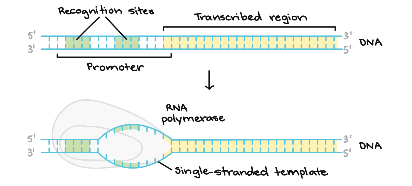
The cell receives an enzyme (a protein) called RNA polymerase, and it is the job of this
molecule to transcribe some DNA into RNA. Polymerase first engages some molecules called
transcription factors to actually find the gene sequence it wants.
Science is still researching why this happens. Genes can be a few thousand up to several
million base-pairs in length. This is called the gene sequence. The gene sequence has what
may loosely be called an address. So the transcription factor finds the beginning of the
gene, and binds the polymerase to the promoter. The promoter sequence is a sequence
preceeding the gene which tells about the gene. It tells if the gene is turned “on”
or “off”,
so is the gene able to produce a protein or not, and it has other coding information.
The polymerase starts its work. First it unzips a short part of the DNA about 20 base-pairs
in length, just like mitosis, by breaking the hydrogen bonds. Then it looks at each base-pair
in turn, and works its way down the DNA sequence, it unzips one pair in front, and rezips
one pair behind. Its job is to read the DNA and produce an exact version as an RNA sequence.
RNA (Ribonucleic Acid), is like one side of the DNA, it is like only one comb if you will.
It has the backbone, and then just the single bases.
As it looks at each base, polymerase grabs the complimentary base (from the cell soup) and adds
it to a sequence of RNA it is making. When it reaches the end of the gene it stops, zips
up the last DNA, releases the RNA sequence it has made, and falls off the DNA.
The “end” of the gene is noted by a special sequence called the “end codon”.
So if the sequence it reads is say CATTGCG, then the compliment of that is GTAACGC,
remember A is compliment of T, and G is compliment of C.
RNA has a “start codon” at one end, and a ”stop codon” at the other end.
This is all happening in the nucleus where the DNA lives, and when the transcription
is completed, then a transport protein carries the new RNA, now called messenger RNA,
or mRNA, along a microtubule and out of the nucleus into the main part of the cell.
Transcription is complete. If the gene sequence was 3,000 base-pairs, then the mRNA is
the same length.
ASIDE There is one small wrinkle however. Transcription does not do the compliment on all
bases. The A base should transcribe to T, but it doesn“t, it transcribes to a new base
called U (uracil). So our sequence from above CATTGCG, actually transcribes to RNA sequence
GUAACGC.
TRANSLATION
Continuing the textbook analogy, if transcription makes a copy of the text, translation
changes the language.
So, now the gene has been converted into mRNA and is in the main body (cytosol) of
the cell.
There is a macro-molecule which is ready to do the next process, it is called a ribosome.
But first we must talk about “amino acids”.
In the human body there are 20 different amino acids. If you do an internet search on
“amino acid” you will again get many hits talking about your diet. The reason for this is
that of the 20 amino acids there are 9 that the body can not make itself and those have to
be ingested from food, these are called “essential amino acids”. But let us assume we have
all the amino acids we need in the cell soup, floating around in the cytosol.
Each amino acid is given one letter of the alphabet. So whilst DNA is C A T G, and RNA is
C A U G, amino acids are A-Z. Well almost, as there are only 20 amino acids.
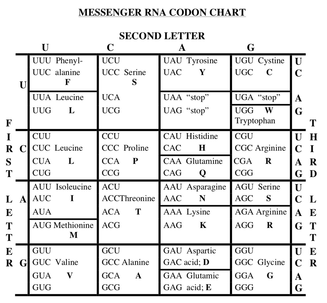
For completeness here is a table of the amino acids, and their diagrams:
Now what the ribosome does with the mRNA is similar to what the RNA polymerase did with
the DNA. It clamps on to the mRNA at the start codon end, and reads down the length,
and this time for every 3 bases (a codon)
it produces one peptide, and adds it to a growing chain called a polypeptide. When it
reaches the end codon it falls off. The mRNA is
retained, and the ribosome may process it again. In fact there may be many ribosomes on the
mRNA strand at the same time. So one strand of mRNA can produce many thousands of protein
parts.
So one peptide is 3 amino acids joined together (a codon). There is a relationship
of all the 3 combinations of RNA to its amino acid:
You don’t need to know all these relationships, except to know that 3 bases
(one codon) code for one amino acid. And in fact three different bases can code for the same
amino acid. For example in the top left corner of the chart, UUU codes for F, and so does
UUC. And in the bottom right corner, GGU GGC GGA GGG all code for G.
UGA is a special codon, as it codes for the “stop” codon, so when the ribosome sees that
it stops translation.
The ribosome also has a special feature. It can error correct. That is, if when it is
reading 3 bases it chooses the wrong amino acid, it can detect its error, go back 3 bases,
and try it again.
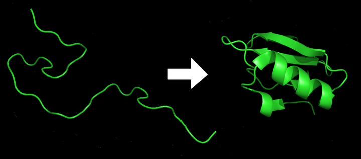
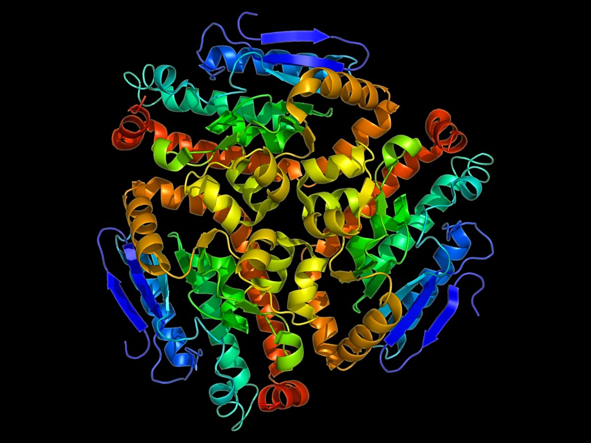
PROTEIN FOLDING
Once the ribosome has fallen off the RNA, it releases the peptide chain. This chain now
starts a remarkable dance, called protein folding. The peptide chain is actually the
protein, but it is not in the final shape.
The chain starts to bend and buckle, it makes and later breaks many hydrogen bonds, and
slowly it starts to make 3 special shapes:
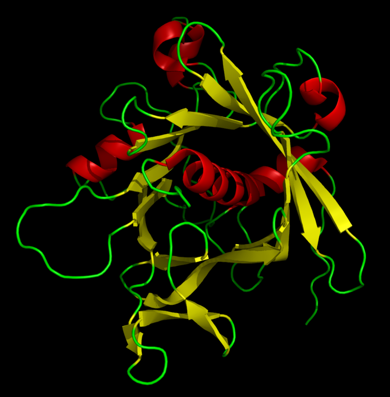
1. alpha helix
2. beta sheet
3. connecting strand
These three shapes will become the building blocks for the next phase.
1. alpha helix This is similar to the DNA shape. The peptide
forms into a spiral. This can vary in length and to how many loops it has.
2. beta sheet. This is a flat long thin structure, a bit like how one would knit a scarf,
zig-zagging as each stitch adds to the width, and then starts a new row to add to the length.
Also this can be any width and any length.
3. connecting strand This is a short length which will act
like a hinge in the next process, and there is a piece between every helix and sheet.
We now have our three building blocks. The protein continues its folding dance,
now using the hinge between each block to twist and turn the sheets and helixes into a new
shape. Again hydrogen bonds are made and broken, thus giving shape to the final protein.
Some proteins are short polypeptides and make a simple protein:
Some proteins are long polypeptides, and make very complex proteins.
The protein is now complete, and if it is destined for the outside world, a
transport protein will grab it, and walk down the microtubule to the membrane and release
the protein out of the cell.
Some proteins are retained inside the cell for its own use.
An example of this is the ribosome itself. The ribosome is actually assembled from several
proteins. So, if the cell is running short of ribosomes it can trigger a polymerase to
start gene expression of the proteins it needs.
The ribosome does have error correction, but mistakes can still be made. Sometimes a protein
can misfold due to outside factors, and this can lead to disease of the host.
HOW PROTEINS WORK
We mentioned that proteins can be characterised into 6 functional types. How they perform
their functions is always the same.
It is the actual shape of the protein which defines it. But also a protein can change
its shape, technically this is called a conformation change. An example of this is the
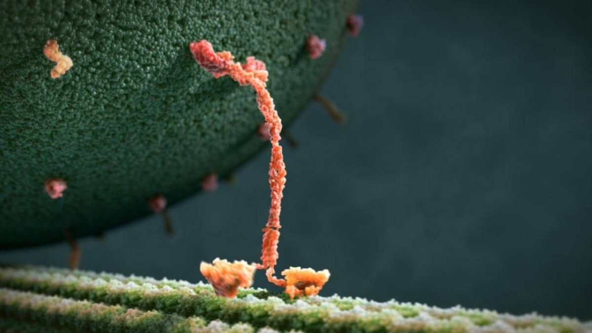
transport protein which walks along a microtubule, actually emulating the human gait. The
video is by Ron Vale.
The orange is the protein (kinesin), at bottom is the microtubule, and the dark
green is the huge cargo. The protein has two “feet”, which alternately grab and release
the microtubule, and flip forward, thus emulating human walking.
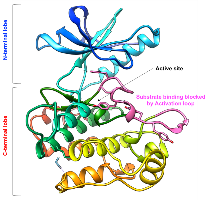
Often proteins have what is called the “active site“or “binding site”.
This is often a
hole in the protein, which allows for other proteins or molecules to bind at the active
site.
The active site is normally a very special shape, thus allowing only a targeted protein or
molecule to bind. A conformational change can inhibit binding. Pharmaceutical drugs
are often designed to fit the active site of an antigen (virus or invader) so stopping it
from being active.
TAKE AWAY
So, the take away from this long and complex chapter are simply this.
a. genes are in the DNA
b. RNA is made from a gene
c. RNA codes for a particular protein.
d. RNA is turned into a protein by a ribosome.
As usual, I am still stunned that all this is able to be done using the 3 main life elements
O H C, and the extra two P N.
7 Viruses
So the question arises, are viruses alive? Well they have DNA or RNA which allows them to
replicate. But they need a host cell to do that. But there are many organisms that have
symbiotic relationships with a host. All humans have hundreds of viruses within them at
any given time, and not all are detrimental, in fact some are considered beneficial.
So, the answer depends on your opinion.
STRUCTURE
Most viruses come with a basic set of structures. They have DNA or RNA which is their
blueprint to replicate called a genome. They have a protective container for that package,
called a capsid.
They may have motility, which means they can move. And they have a mechanism to breach the
cell wall and deposit their genome package inside.
Viruses are made of several proteins, depending on their type.
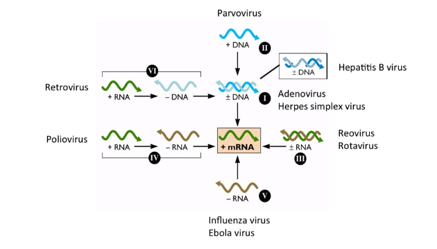
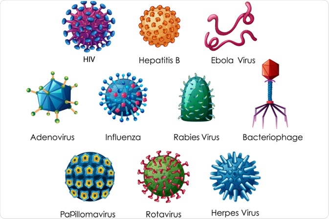
TYPES
There are considered to be 7 different types of virus, depending on their genome, and
their shape. The genome may be DNA, broken DNA, circular DNA, +RNA, -RNA and several others.
These shapes have been defined in a system called the Baltimore Classification.
The chart shows 6 types, but type 6 is now considered to be 2 types.
The genome can be any of a variety of types not found in other organisms. Their shape also
can vary a lot.
REPRODUCTION
The basic method of reproduction, is for the virus to inject their genome into the cell.
From there it is a simple matter of letting the ribosomes do what they do. If the virus
is of a DNA type, it will normally have a polymerase of its own included, and the DNA
will be turned into mRNA. If the virus is of an RNA type, then no action is required.
The ribosome does not know the difference between mRNA from its own cell or a virus, and
just makes the virus proteins. Coronavirus has 8 pieces of RNA, and these code for the
proteins that make the capsid, the famous spike and other components.
Once the ribosome has made all the proteins, one of them replicates the genome, another
assembles the capsid and puts the new genome inside, and assembles the rest of the virus.
Then a transport protein moves the virus to the cell wall, and releases it through.
Some viruses don‘t release the virus, and the numbers grow in the cell and the pressure
increases until the cell wall finally bursts, releasing millions of virus particles out.
8 Immune System
The immune system is considered to be in two parts, the innate system and the adaptive system.
Simply put the innate system is what is always there, it never changes and depends effectively
on physical blocks to invasion by antigens. The adaptive system works on the idea that once
an invasion happens it will start working to fight that particular antigen.
INNATE SYSTEM
This part of the immune system are the physical barriers, and are the ones you were born with.
These include the skin, sweat, tears, mucus, stomach acid, and the cough response.
ADAPTIVE SYSTEM
This part of the immune system is the really complicated part. It takes a few days for this
system to mount a defence to an antigen during which time the antigen builds a stronghold,
and once the adaptive system starts then the war begins.
Once the war begins it becomes a statistical battle, as to which side has the greater
number of fighters.
The adaptive system has two main types of soldiers.
B-CELLS, T-CELLS
The job of B-cells is to patrol the body looking for antigens (invaders like viruses
and microbes). It has one main weapon. Every cell in the host body has a special tag on
the outside of the cell, we call this the SELF tag. So the immature (naive) B-cell
interrogates every cell it meets “are you SELF?”, if yes, the B-cell moves on, if not
it has met NON-SELF, an antigen.
It takes a small “bite” out of the antigen, normally one of the proteins that comprise the
antigen body. It absorbes the bit and reshapes it a little and then presents it on the outside
of its cell wall as a tag. The B-cell is now considered “mature”, and its job changes.
It is now forever related to that antigen, and starts to replicate itself (mitosis)
including the tag, millions of times. Some of those become antibodies, which will target
that specific antigen.
The B-cell also makes friends with a T-cell, and gives it the same tag. These T-cells are
also known as killer-T-cells, and they go off looking for host cells that have become
infected.
When a host cell becomes infected it puts a tag outside its cell wall as a cry for help,
and also releases a bundle of little communication proteins called cytokines.
The cytokines attract more B-cells to help.
Also the killer-T-cells read the tag outside the infected cell, and instigate cell death
(apoptosis) and kills the cell, and everything in it.
And so this war continues, until eventually the host wins. The T-cells reduce, and so do
the B-cells. However a bundle of the B-cells remain, and are called memory-B-cells. Memory
B-cells remain floating around the host, ready to react if the same antigen ever invades
again. If there is another infection of the same antigen, memory-B-cells are able to react,
and do so much quicker this time.
And it is this feature of the adaptive immune system that is used by vaccines.
9 Vaccines HOW DO THEY WORK
Modern vaccines come from a discovery in 1796 when a doctor (Edward Jenner) noticed
that milkmaids who got cowpox from the cows were immune to the much more serious disease
smallpox, which was decimating Europe at the time. He took a small swab of a milkmaid’s
cowpox sore, and injected it into his son, who escaped the ravages of smallpox. It was a
terrible risk, but it worked.
So, vaccines work on the idea that a small non-lethal dose of the disease will protect
against the full disease in future.
Effectively what is happening is that the initial application of the small dose causes
the adaptive immune system B-cells to react, and overcome the dose, leaving memory-B-cells
behind for that disease.
ATTENUATED LIVE VACCINE
Modern vaccines take the real virus, and attenuate it. The process of attenuation requires
injecting mice with the virus, and those that survive have their blood plasma injected into
other mice, and those that survive…etc.
So after many cycles of reinfection, the blood plasma still has the virus, but it is
attenuated, meaning it has less or no effect.
Other modern techniques of attenuation involve modifying the genome of the virus, to make
a vaccine.
In the early days of virology, however, the process was still not exact. In 1955, 200,000
children in USA received polio inoculation, but the vaccine was defective, and 40,000 were
infected with polio. Ten died and many were paralized. This "Cutter Incident" led to
improved techniques of attenuation.
ASIDE: By now the polio virus has been all but eradicated. Only Pakistan
and Afghanistan have any polio left in all of the whole world.
mRNA VACCINE
This type of vaccine works in the same way that viruses do. The idea is to introduce some
selected mRNA into the cell and allow the ribosomes to create a protein which looks like
part of the antigen (virus) of interest.
When that protein is released, the B-cells see it as NON-SELF, go through their usual
process, although there is not really all out war. What remains are the memory-B-cells.
The host is now prepared for an actual infection of the antigen.
The mRNA vaccine idea was 15 years in the making. As I mentioned earlier, DNA is a robust
molecule, whereas RNA is a lot less so. So the idea of using a small part of the virus RNA
was valid but the vector (how you transmit the package) was more difficult. The vector
where you take a virus, say adenovirus, remove the package, and put in what you want,
was OK, but many people have B-cells for that and other viruses, so the vaccine would
be attacked immediately. Finally around 2019, just as the Coronovirus Pandemic was
starting a technique was perfected.
The RNA was wrapped in a coat of lipids. Lipids are molecules that hate water, and clump
together. So, the mRNA was wrapped in a lipid bundle (called a nano-particle), and injected
into the host. A nano-particle is any small particle, like a protein, or indeed a virus.
The cell membrane accepted the lipid bundle and the RNA entered the cell. The ribosomes do the rest.
10 Coronavirus
Coronavirus is a name given to a group of viruses, which include SARS-CoV-2, MERS-CoV,
SARS-CoV (the original which has been backnamed to SARS-CoV-1), HKU1, and several others,
including the common cold.
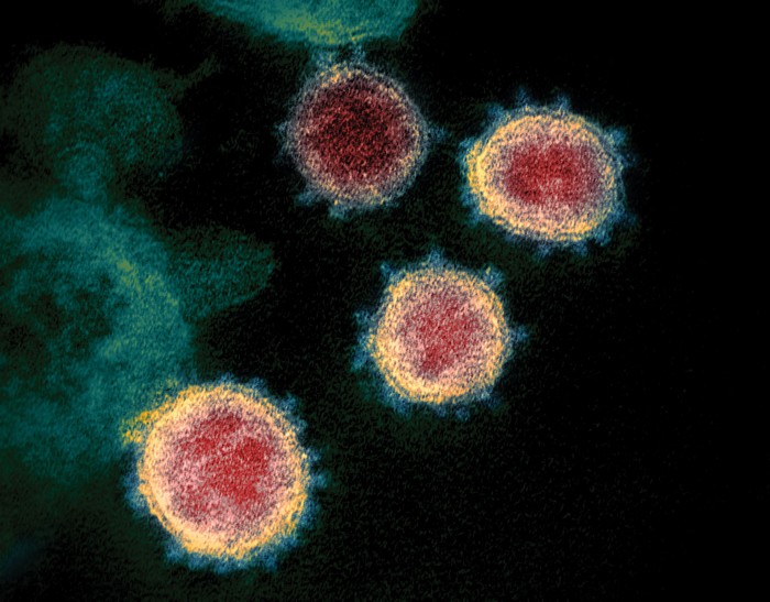
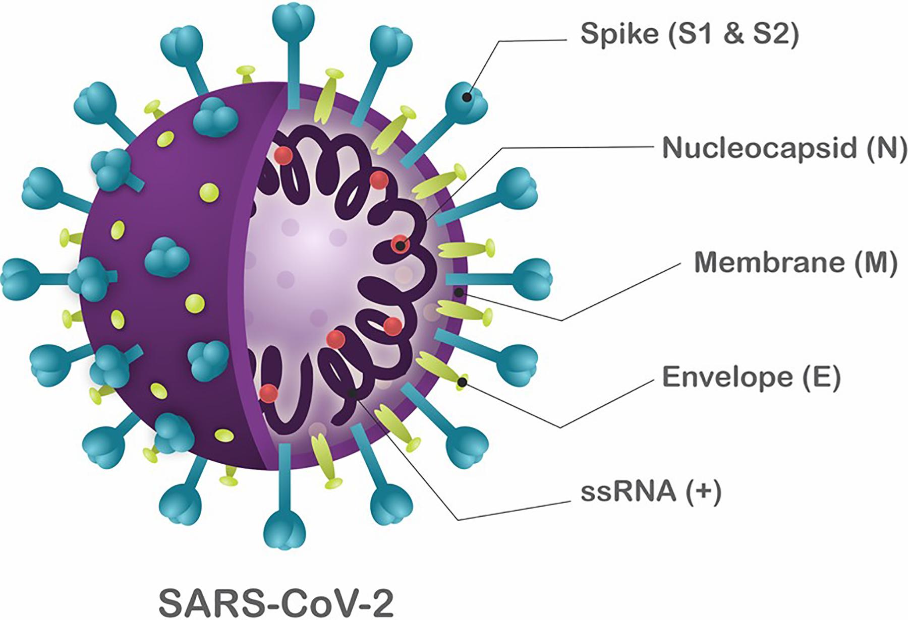
Here is a picture of an actual coronavirus. All images in this document are diagrams of
what scientists think things look like, this is a picture, made with a light microscope.
It has been colour enhanced. To it's right it is in diagram form.
Coronavirus is an RNA virus with 8 pieces of RNA and 29,881 base-pairs in length,
using 9,860 amino acids to make 29 proteins.
The virus is airborne, which means that infection of a host is accomplished by an infected
person coughing or sneezing, the virus rides the air in tiny mucus droplets, is breathed
in by the host, and the virus infects the soft tissue of the upper respiratory tract.
The virus bypasses the innate immune system, and is able to infect the cells of the throat
which are an easy target.
The virus cannot survive outside a host for very long, and there is no evidence to suggest
it can infect a host from touching a surface. It may however survive on a surface for a
short time, be picked up on a person's hand, and infect the host if the person touches their
eye, or licks their fingers.
CELL ENTRY
On the outside of
many cells are receptors. These are proteins that have a binding mechanism that allows
a protein of the correct shape to open a hole in the cell wall and inject molecules that
the cell may need. There is one such receptor called ACE2 (angiotensin-converting enzyme 2),
which allows molecules into the cell that help control the blood pressure of the host.
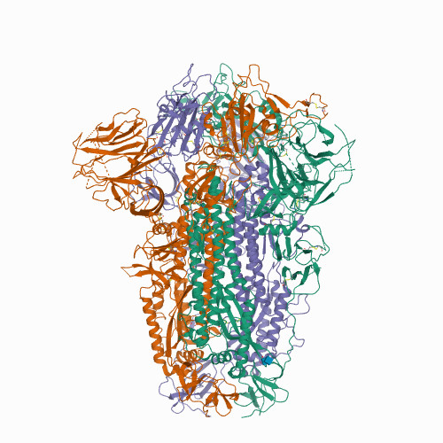
Coronavirus has adapted its spike protein to use that receptor for its own use, which is
to open the cell wall to inject its genome. Many viruses have adapted this technique of
using a receptor that is designed for something else. Here is a diagram of the spike.
The spike connects to the ACE2 receptor, it does a conformation change to open
a hole and inject its genome package into the cell.
COVID-19 DISEASE
The disease coronavirus (SARS-CoV-2) causes is called CoViD-19, “corona virus disease”
first seen in 2019.
The disease is a form of pneumonia, and as with that condition, death is the result of
the person effectively drowning in their excessive phlegm. Treatment often involves
administering high levels of O2, but that has had limited success.
Poor outcomes often happen to patients with comorbidity. This is where a patient has
two or more diseases at the same time. The elderly also have a higher risk of poor
outcomes.
The disease can be made worse by a condition called “cytokine storm”. Cytokines are a
cells method of drawing attention to the fact that it is stressed. The stress can be an
infection or a physical assault, like a cut. The storm happens when the cells release too
many cytokines too quickly, and once it starts, there is a positive feedback which causes
other cells to do the same.
Early in the infection, coronavirus tickles the upper respiratory tract, and makes the host
cough and sneeze. Thus the virus is thrown out into the air, and is ready to infect a new host.
So you see a host can be infected by many viruses and even variants of the same virus.
VARIANTS
When a virus particle enters the cell, and the ribosome does its work, one protein it
creates does the virus RNA replication, and puts it in the virus capsid. When the replication
happens, sometimes the virus RNA is not copied properly.
Some call this a mutation, but I like to call it a simple change.
If that change makes the virus a better fit in the soup, then it will replicate more
than other virus versions, and if it replicates in many hosts, scientists then refer to it
as a variant.
Historically viruses were named for the place where they were discovered. The 1918 flu
pandemic was initially called “Spanish Flu”. But in recent times scientists thought it was unfair
to call a virus by a place name, and they
started to use the Greek alphabet, hence the variants, beta and gamma and omicron.
It is possible for a host to have many viruses at the same time, and many changes in each
virus.
The tests to see if a host is infected with the SARS-CoV-2 antigen fall into
two categories,
a full test to find the virus DNA/RNA, and a virus protein test which is quicker but less accurate.
PCR TEST
Before we talk about the PCR Test, we need to explain what PCR itself is. The scientist who
invented the PCR procedure won the Nobel Prize for his efforts.
When dealing with DNA, the samples are often extremely small, with a low strand count. In
fact such a low strand count that it is difficult, if not impossible, to detect them. So a
technique to multiply the count of a sample was invented called Polymerase Chain Reaction (PCR).
The process is similar to mitosis in cells, but performed on a sample in the lab.
First the sample is heated, and this denatures (unzips) the DNA. Then a polymerase is
introduced with a solution of C A T G, and this completes the unzipped DNA. This has doubled
the strand count.
Then the process is repeated, with the strand count doubling every cycle.
Often the cycle is repeated to a count of 40.
A coronavirus PCR Test is as follows. Coronavirus is an RNA virus so the first step is to
“Reverse Transcriptase” to turn the RNA into DNA.
You remember in The Central Dogma,
Transcriptase turns the DNA of a gene into mRNA, well this is the reverse of that, hence its name.
Once we have a DNA version, then PCR cycles are applied.
Now the count becomes important. Each cycle doubles the strand count, and after each cycle
a test is done to see if DNA is detected. If the count gets to say 25, and DNA is detected,
then the test is positive. If the count gets to 30 and no DNA is detected the test is
considered negative.
When first invented, these tests were extremely labour intensive, and took several weeks.
Now larger PCR machines can process over 1500 samples per batch, in less than a day, and
these machines can cost as little as $10,000.
RAT
Rapid Antigen Test. PCR Test detects the DNA/RNA of the virus is present. The RAT detects
if a particular virus protein is present, a bit like what a B-cell does. These tests are
fast (20 mins), and cheap ($20), and can be done by a patient at home.
The test unit has an enzyme in it which binds to the active site of a chosen virus protein.
The enzyme has a fluorescing molecule attached to it, so if the virus protein is present in
numbers, then the enzyme will bind to them all and light up. It will light up sufficiently
to be seen by eye, and that is considered a positive test.
This test is fast and cheap, but the chance of a false positive, or false negative is higher.
EXPERIMENTS
There are hundreds of thousands of virologists and immunologists who have been working
diligently, and their work has been viewed in more detail since the COVID-19 pandemic.
They fall into two funding methods, one is the academic where labs are funded by open grant
applications, and the others are funded by pharmaceutical companies in secret. I will not
discuss the latter.
There is an old academic adage - “Publish or die”. This means you must do research and
publish the results or have your career wilt. But the papers that are published are the
most important component of scientific research.
So what happens is that a professor at a university has an idea, applies for and is given
grant money. She fills a lab with equipment, and populates the lab with undergrad students,
who will do all the work under her guidance. Many professors travel the world visiting
symposiums, where they engage with others working in similar fields. Often they will be
working on the same thing, and then start a collaboration. So a US lab could collaborate
with similar labs from UK, EU and maybe China, politics and culture play no role.
At the appropriate time they will publish a paper on their work. The “Scientific Paper”
is the most important output from research.
At first it is loaded onto a web Database, and this is called a “pre-print”, because it
is yet to be peer-reviewed. The review process is one where many interested scientists
look at the paper and critique it. Once that is done, and changes are made, then the paper
can be printed, which means it can be released to scientific magazines. In virology the
most prestigious are “Cell” and “Nature”. The final published
paper overwrites the pre-print in the online databases.
When a professor starts a project, and indeed all through its work, she will do a search
of the databases to see the state of the field. Was it Einstein who may have said he stood
on the shoulders of giants? Well that is what is happening here. Every scientific paper
moves the knowledge forward by a tiny bit. And so the research inches forward, with each
new move published in a paper.
Within the paper the work is explained in exact detail, in language only a similar scientist
will understand. It must be exact. Normally the paper will describe one or more experiments
that were performed, and the exact results obtained.
In virology many of the experiments are performed in a lab, on samples of interest in petri
dishes. Some of the pathogens are extremely dangerous, like for example ebola. So each lab
is rated for its “Bio Safety”, and gets a BSL number. BSL-1 is the least dangerous, and work
is done in lab coats. BSL-4 is the most dangerous, and work is done in secure labs, with
scientists wearing special suits that make them look like balloons.
Experiments are done according to the
“Scientific Method”, which is a set of agreed rules,
the most important of which is that the experiment MUST be
repeatable anywhere in the world.
The experiments have developed over time, and as scientific technology has developed.
The simplest experiments involve putting a sample in a petri dish, and adding some agent
to see what happens. The sample is reviewed every day to see if the colony grows or dies.
This technique is fine for microbes and macro-molecules, but to research viruses a more
detailed approach is needed, and that is “X-ray Crystallography”.
This is where a sample is embedded in a crystal thus freezing it in time. Then the crystal
structure in bombarded with X-rays. These sometime hit an atom’s nucleus and are deflected.
Behind the crystal sample are sensors which record where the X-rays end up. Computers look
where the Xw-rays come from, and where they end up, and build a 3-D picture of the sample.
So, in this way the position and type of every atom in the sample is identified.
This web page is entitled “From Atoms to Viruses”, and I suggest we have now come full
circle, as at the end the research into viruses often entails identifying the exact atom,
its position, and its covalent bonds.
When this technique was first invented it took a scientist up to a year to create a viable
crystal, and only now are computers good enough to make the ribbon diagrams of proteins we
saw earlier.
Also, if there is a conformational change in a protein, the scientist has to make and
anaylze the crystal before and after the change. This is hard, detailed and exact work.
X-ray Crystallography, is now being superseded by a new technique called
“Cryogenic Electron
Microscopy”, or cryo-EM for short. X-rays are replaced by electron beams, and the crystal is
replaced with very low temperature samples (the cryo in the name). This new technique allows
for much quicker turn-around, and removes the need for the difficult process of making a
crystal. There is even talk of “Video cryo-EM”, which would allow movement to be checked.
TRIALS
The research done by academia pushes forward knowledge of the subject. The research done
by big pharma companies is to make drugs to help people…well and to make a lot of money.
An old adage is that the first pill costs $5,000,000 and every pill after that costs $1.
The first time a real biological entity is trialled on, is normally the mice in the academic’s
lab. Rodents make up 40% of mammals on earth, they are hardy, easy to work with and cheap.
Sometimes a lab will work with what they call non-human primates, but these can cost
$40,000 each. And sadly at the end of each experiment, the animal is “sacrificed”, and an
autopsy is done.
Once the animal trials are completed to the scientists satisfaction, and a promising drug
that worked in the animal model, it is time for clinical human trials.
There are 4 types of human clinical trials, called “Phases”.
Phase-1 trial involves a small group of 20-80 persons. This is a safety check, and dosage
review.
Phase-2 trial involves several hundred. This is a review of efficacy, that is, does it work?
Phase-3 trial can involve up to 100,000 people. This is the final study before approval
for general use.
Phase-4 trial involves review of the drug in general use over many years.
The Phase-4 trials actually fall into the domain of immunology, which is the big-picture
research of large data-sets, to see what is really happening to a whole population, like
is being done with the COVID-19 pandemic, to watch how it is progressing.
13 Conclusion
So, I hope this VERY simplified journey from Atoms to Viruses was of interest.
I am still surprised that almost all the chemistry is done on the 3 main atoms with a small
few assistants.
I am further surprised (always), that scientists can find out so much about a world that
can not even be seen by humans.
I want to thank Prof Vincent Racaniello of Columbia University, USA. In
January 2020, just
as the COVID-19 pandemic was starting, I found his Virology lectures online. The course
lasted 20 weeks, but it took me 18 months, as I had to learn basic chemistry (a subject I
hated at school). But now at age 71, I am finally catching up, and I find life chemistry
fascinating. At my age, and location (Perth, Australia) and with travel restrictions in
place, I am sad I will not be able to travel to USA to visit so many of the places that
have become so central to the study of virology.
They don’t know it, but I thank all the researchers that ever handled a pipette.
With the now invented mRNA vaccines, I see a door opening. This technique allows for the
scientists to create ANY protein they want, and I see this as a new treatment technique,
which may revolutionise healthcare.
There are many in the world who see this as the beginning of nano-bots invading our bodies,
of a malicious scientist taking over the world. Many new technologies we now call normal
were once viewed in this way, and yes there will always be risks. I think of Dr. Jenner
injecting his own son with live pox. But we live in a world of fantastic advancements,
and I think if we take care, have oversight, and are mostly brave, we will benefit hugely.
If you own any of the images included in this web page, I would
like to give you credit, or if you object I will remove them, please contact
rikf.a2v@gmail.com